
 31 Mar 2022
Gastroesophageal reflux disease (GERD)
Diagnose
Your doctor may be able to diagnose GERD based on a physical exam and a history of your signs and symptoms.
To confirm a diagnosis of the GERD, or to check for complications, your doctor may recommend:
1. Upper endoscopy
Your doctor inserts a thin, flexible tube with a light and camera (endoscope) down your throat to examine the inside of your esophagus and stomach. Test results are often normal when reflux is present, but the endoscopy may reveal inflammation of the esophagus (esophagitis) or other complications. An endoscopy may also be used to collect a sample of tissue (biopsy) to be examined for complications such as Barrett’s esophagus.
2. Mobile acid (pH) probe test
A device is placed in the esophagus to determine when and for how long stomach acid will reflux there. The monitor connects to a small computer that you wear around your waist or a strap on your shoulder. The monitor may be a thin, flexible tube (catheter) passed through your nose into the esophagus, or a clip that is placed into the esophagus during endoscopy and passed in the stool about two days later.
3. Esophageal manometry
This test measures regular muscle contractions in the esophagus when swallowing. Esophageal manometry also measures the coordination and force exerted by the esophageal muscles.
4. X-ray of the upper gastrointestinal tract
An X-ray is taken after you drink a chalky liquid that coats and fills the inner lining of your digestive system. The wrap allows your doctor to see the silhouette of your esophagus, stomach, and upper intestine. You may also be asked to swallow a barium pill, which can help diagnose esophageal strictures that may interfere with swallowing.
Treatment
Your doctor will likely recommend that you try lifestyle modifications and take over-the-counter medications first. If you do not feel relief within a few weeks, your doctor may recommend a prescription or surgery.
Over-the-counter drugs
Options include:
1. Antacids that neutralize stomach acid
Antacids, such as Mylanta, Rolide, and Toms, may provide quick relief. But the antacids alone won’t heal an inflamed esophagus damaged by stomach acid. Overusing some antacids can sometimes cause side effects, such as diarrhea or kidney problems.
2. Medicines to reduce acid production
These drugs – known as H-2 receptor blockers – include cimetidine (Tagamet HB), famotidine (Pepcid AC) and nizatidine (Axide AR). H-2 receptor blockers do not work as quickly as antacids, but they provide longer relief and may reduce acid production from the stomach for up to 12 hours. Stronger versions are available by prescription.
3. Medicines that block acid production and treat the esophagus
These medications known as the proton pump inhibitors are stronger acid blockers than H-2 receptor blockers and allow time for damaged esophageal tissue to heal. Over-the-counter proton pump inhibitors include lansoprazole (Prevacid 24hr) and omeprazole (Prilosec OTC, Zegerid OTC).
Medicines
Strong prescription treatments for GERD include:
1. Strong prescription H-2 receptor blockers
These medications include famotidine (Pepcid) and nizatidine. These medications are generally well tolerated but their long-term use may be associated with a slightly increased risk of vitamin B12 deficiency and bone fractures.
2. Strong prescription proton pump inhibitors
These medications include esomeprazole (Nexium), lansoprazole (Prevacid), omeprazole (Prilosec, Zegerid), pantoprazole (Protonix), rabeprazole (Aciphex) and dexlansoprazole (Dexilant). Although these medications are generally well tolerated, they may cause diarrhea, headache, nausea, and vitamin B12 deficiency. Chronic use may increase the risk of developing a hip fracture.
3. Medication to strengthen the lower esophageal sphincter
Baclofen may relieve GERD by decreasing the frequency of relaxation of the lower esophageal sphincter. Side effects may include the fatigue or nausea.
Surgery and other procedures
Gastroesophageal reflux disease can be controlled with medication. But if medications don’t help or you want to avoid long-term use of medications, your doctor may recommend:
1. Fundoplication
The surgeon wraps the upper part of your stomach around the lower esophageal sphincter to tighten the muscles and prevent GERD. Fundoplication is usually done through a minimally invasive (laparoscopic) procedure. The bypass of the upper part of the stomach can be partial or complete.
2. LINX device
A ring of the tiny magnetic beads is wrapped around the junction of the stomach and esophagus. The magnetic attraction between the grains is strong enough to keep the junction closed to reflux acid, but weak enough to allow food to pass through. The LINX can be implanted using minimally invasive surgery.
3. Transoral fundoplication (TIF)
This new procedure involves tightening the lower esophageal sphincter by making a partial wrap around the lower esophagus using polypropylene stabilizers. TIF is performed through the mouth with a device called an endoscope and does not require a surgical incision. Its advantages include the fast recovery time and high tolerance.
If you have a large hiatal hernia, the TIF alone is not an option. However, it may be the possible if TIF is combined with laparoscopic hiatal hernia repair.
BY: Web Team
Magazine
COMMENTS: No Comments
 31 Mar 2022
Neuroendocrine Tumors: Types of Treatment
This section explains the types of treatments that are considered the network’s standard of care. “Standard of care” refers to the best known treatment, which is usually based on strong evidence from previous clinical trials. When making decisions about a treatment plan, we also encourage you to consider clinical trials as an option. A clinical trial is the research study that tests a new approach to the treatment. Doctors want to know if the new treatment is safe, effective, and possibly better than the standard treatment. Clinical trials can test a new drug, a new combination of standard treatments, or new doses of standard medications or other treatments. Clinical trials are an option for treatment and care for all stages of cancer. Your doctor can help you think about all of your treatment options.
How is NET treated?
In cancer care, different types of doctors often work together to create a comprehensive treatment plan for the patient that combines different types of treatments. This is called a multidisciplinary team and is especially important for people with .NET. Cancer care teams include a variety of other healthcare professionals, such as physician assistants, nurse practitioners, oncology nurses, social workers, pharmacists, counselors, dieticians, and more.
Treatment is the options and recommendations depend on several factors, including:
- Primary location (where the network originated)
- Whether the tumor is functional or not
- The stage (where the tumor is in the body)
- Degree and degree of differentiation (how quickly cells divide)
- growth rate
- Somatostatin receptor status (whether tumor is bright on 68Ga DOTATATE PET)
- Possible side effects
What is the patient’s preference and general health?
Take time to learn about all treatment options and be sure to ask questions about things that aren’t clear. Talk with your doctor about the goals of each of the treatment and what you can expect while receiving the treatment. These types of conversations are called “joint decision making.” Decision-making is involved when you and your doctor’s work together to choose treatments that best fit the goals of your care. Joint decision making is especially important to the network because there are different treatment options. Learn more about making treatment decisions.
The common types of net treatments used are described below. Your care plan also includes treatment for symptoms and side effects, which are an important part of cancer care. Visit the individual section for a specific type of NET for more information about treatment options (see Introduction for a complete list).
Active monitoring
Sometimes, active monitoring may be recommended. This method is also called watchful waiting or watch and wait. It is used most often for someone with low-grade NET that may grow slowly and not spread or cause problems for months or years. Effective treatment usually begins if the tumor shows signs of growth or spread. With this approach, the tumor is closely monitored with the regular tests, which may include:
Imaging the tests, usually CT scans or sometimes MRI scans (see Diagnosis)
Blood tests
Physical examinations
Surgery
Surgery is the removal of the tumor and some surrounding healthy tissue during the operation. A surgical oncologist is a doctor who specializes in the cancer surgery. Removing the entire tumor is the standard treatment, when its possible. Most in situ meshes are successfully treated with surgery alone. The surgeon usually removes some of the tissue surrounding the tumor, called the margin, in an effort to leave no trace of the cancer in the body.
When the tumor cannot be completely removed, sometimes “reduction surgery” is recommended. Volume removal surgery removes as much of the tumor as possible and may provide some symptom relief, but it generally does not cure the NET.
People who have had carcinoid syndrome are at risk of developing a carcinoid crisis during surgery (see Symptoms and Signs). To avoid major complications from a precancerous crisis, the anesthesiology team must be fully aware of this risk prior to surgery, so that they can have treatment on hand to control symptoms. Octreotide is usually given into a vein before surgery to prevent a cancerous attack.
Before surgery, talk with your health care team about possible side effects from the specific surgery you’ll have.
If the NET cannot be removed with surgery, the tumor is called “non-operative.” In these cases, the doctor will recommend another treatment plan.
Treatments with medication
Treatments using drugs are used to destroy cancer cells. The drug can be given through the bloodstream to reach cancer cells throughout the body. When medication is given in this way, it is called systemic therapy. The drug can also be given locally, which is when the drug is applied directly to the cancer or kept in one part of the body.
Targeted therapy
Targeted therapy is treatment that targets genes or proteins specific to the tumor or tissue environment that contribute to cancer growth and survival. This type of treatment prevents the growth and spread of cancer cells and limits damage to healthy cells.
Not all tumors have the same goals. To find the most effective treatment, your doctor may run tests to identify genes, proteins and other factors in the tumor. This helps the doctors better match each patient with the most effective treatment whenever possible. In addition, the research studies continue to discover more about the specific molecular targets and new treatments directed at them. Learn more about the basics of targeted therapies. Talk to your doctor about the potential side effects of a particular medication and how they can be managed.
Everolimus (Afinitor) is a targeted therapy approved by the US Food and Drug Administration (FDA) for the treatment of advanced NETs of the gastrointestinal tract, lung, and pancreas. This drug targets a protein called the mTOR that is important for the cell growth and survival. This medication can help slow the growth of these tumors in some patients, but it does not usually shrink the tumors. Side effects include mouth sores, low blood counts, and fatigue.
BY: Web Team
Magazine
COMMENTS: No Comments
 31 Mar 2022
Reproductive Endocrinology
Reproductive endocrinology is a subspecialty of obstetrics and gynecology (OB/GYN) that focuses on the evaluation and treatment of hormonal disorders, anatomical disorders, and other conditions that can affect fertility (the ability to become pregnant) or the ability to carry a pregnancy to term. Doctors who practice in this field of OB/GYN are called reproductive endocrinologists. Reproductive endocrinologists first complete training in obstetrics and gynecology before completing advanced training in reproductive endocrinology.
Reproductive endocrinologists treat many problems involving the reproductive organs or affecting pregnancy and childbearing, including:
- Infertility in women and men
- recurrent pregnancy loss
- Menstrual disorders
- uterine fibroids
- Endometrial
- Polycystic ovary syndrome
- early menopause
- Early or late puberty
- Congenital defects of the genitals
- Hormonal disorders that affect reproduction
Most reproductive endocrinologists focus their practice on fertility issues. As such, their goal is to improve and maintain fertility potential (in both men and women) and to identify and treat the causes of infertility.
An obstetrician-gynecologist or primary care physician may refer a woman or couples to a reproductive endocrinologist after failed attempts to conceive. In general, further evaluation by a reproductive endocrinologist is warranted if a couple has not succeeded in conceiving after 12 months of unprotected intercourse if the woman is younger than 35, or after 6 months of unprotected intercourse if the woman is older than 35 years old.
An infertility assessment begins with a thorough physical examination and a detailed personal and family medical history for each partner. This is followed by a careful review of the results of blood tests, semen analysis, and imaging tests to look for an anatomic cause of infertility (eg, a problem with the uterus, ovaries, or fallopian tubes).
Once the diagnosis is established, an individualized treatment plan is provided, with the ultimate goal of a successful pregnancy. Treatments may include monitoring hormone levels, giving medications to stimulate ovulation or regulating hormones, or surgically correcting a problem that interferes with conception (for example, a blocked fallopian tube).
If pregnancy cannot be achieved with these measures, reproductive endocrinologists are skilled in assisted reproductive techniques, such as:
- Intrauterine insemination – the introduction of sperm directly into the uterus to facilitate fertilization
- In vitro fertilization (IVF) – the fusion of an egg with semen outside the body in a laboratory for fertilization, then transfer of the fertilized egg (embryo) to the uterus
- Intracytoplasmic sperm injection (ICSI) – similar to ICSI, except that a single sperm is injected directly into an egg in a lab, then the embryo is transferred to the uterus
- Egg or Embryo Cryopreservation – Freezing of an Egg or Embryo for Future Pregnancy Attempts
- Sperm Freezing – Freezing sperm cells to preserve them for future pregnancy attempts
- Ovarian tissue cryopreservation – a procedure to preserve a portion of the ovary for future pregnancy attempts (usually for women whose fertility is threatened by cancer)
- Once a woman becomes pregnant, medical care is often transferred to the obstetrician-gynecologist, who will continue to monitor the pregnancy until the baby is born.
Reproductive endocrinologists are trained in minimally invasive and open surgical techniques to help treat a range of reproductive organ abnormalities. For example, they can perform surgery to remove uterine fibroids, uterine polyps, ovarian cysts, or scar tissue, treat endometriosis, correct congenital abnormalities, open blocked fallopian tubes, and tie off varicose veins in the testicles.
Reproductive Endocrinology and Thumbay University Hospital
Thumbay University Hospital offers expert care through a multidisciplinary team of compassionate reproductive and caring professionals, including geneticists, reproductive endocrinologists, perinatal specialists, diagnostic experts, nursing staff and highly trained technicians. Our team will assist you throughout your personal experience from diagnosis to your individual treatment plan.
BY: Web Team
Magazine
COMMENTS: No Comments
 31 Mar 2022
Treatments for sexually transmitted diseases
Don’t try to cure yourself of a sexually transmitted disease. These diseases are contagious and dangerous. You must see a doctor.
STIs can be treated with antibiotics if treatment is started early enough. STIs cannot be cured, but you can control symptoms with medication. There is a hepatitis B vaccine, but it won’t help if you’re already infected.
If you have been given antibiotics to treat an STD, it is important that you take all of the medicines prescribed to you, even if your symptoms disappear. Also, do not take someone else’s medication to treat your injury; It may make it more difficult to treat.
Here are some specific treatments for STDs:
1. HIV/AIDS
Because AIDS is incurable, treatment focuses on keeping HIV levels under control. Antiretroviral medicines are the standard treatment for HIV infection, and you will usually be given several medicines to take, which is called a ‘cocktail’. The question of when to start antiretroviral therapy for HIV remains a matter of debate. Some doctors believe there is an early start to better manage HIV, while others believe it is best to wait because medications can cause unpleasant side effects and drug resistance may develop. Talk to your doctor about when to start antiretroviral therapy.
2. Chlamydia and the gonorrhea
These STDs are treated with antibiotics. You should start taking it if tests show you have chlamydia or gonorrhea or if you have been exposed to them, even though you may not have symptoms. Your sexual partners will also need to be treated regardless of whether or not they have symptoms. Certain strains of gonorrhea have become resistant to some antibiotics, so you may have to take more than one medication to combat gonorrhea. Make sure your partner also seeks treatment. The test should be repeated after three months to make sure the infection has cleared, even if your partner has been treated. Failure to treat chlamydia or gonorrhea can result in permanent damage to the reproductive organs and the inability to become pregnant.
3. Syphilis
Penicillin is the preferred treatment for syphilis. Early treatment is critical to prevent bacteria from spreading and damaging other organs.
Genital herpes: Once you have genital herpes, the virus remains in your body for life. After the first outbreak, herpes may flare up several times a year, but these episodes may lessen over time. Antiviral medications (such as Famvir, Valtrex, and Zovirax) can help reduce the length and severity of initial and subsequent herpes outbreaks. If you have frequent outbreaks, you may want to use suppressive therapy. In suppressive therapy, your doctor prescribes medication to take every day, to prevent herpes outbreaks.
4. Genital warts
There is no standard for treating genital warts. Most genital warts go away without treatment, so your doctor may choose to do nothing. However, you will still carry the virus that causes warts and can still pass it on to your sex partners. If you choose to treat genital warts, you have several options. Freezing warts or applying medication directly to them is often the first option. If the genital warts do not respond to these options, surgery may be the necessary to remove them. Keep in mind that treatment does not get rid of the infection, and you can still pass it on to others.
5. Hepatitis B
The goal of hepatitis B treatment is to stop liver damage by preventing the spread of the virus. There are now five drugs approved for the use in hepatitis B: adefovir, entecavir, interferon alpha, lamivudine, and pegylated interferon. Each has its pros and cons that you should discuss with your doctor. If you develop the significant liver damage from the hepatitis B, a liver transplant may be necessary.
6. Trichomoniasis
Infection with this organism is treated with metronidazole, and the cure rate is about 90%. If you are pregnant, you can take the medicine. Your partner should also be treated. The test is repeated after three months to ensure that the infection has cleared. Do this even if your partner has also been treated.
BY: Web Team
Magazine
COMMENTS: No Comments
 31 Mar 2022
Skin biopsies: what to expect
When you notice a rash or a worrisome mole on your skin, which is the largest organ in your body, it’s a good idea to see a dermatologist for an evaluation. Sometimes after examining the area, a dermatologist may recommend a skin biopsy.
Skin biopsies are an important part of verifying the diagnosis. For example, a biopsy is the only way for a doctor to confirm and determine the severity of melanoma – the most dangerous type of skin cancer. A skin biopsy may also be used to confirm that a skin tumor is benign or to diagnose inflammatory skin conditions such as drug-related rash or eczema.
Here are the Quick 5 steps involved:
1. A dermatologist examines your skin
Patients see dermatologists for a variety of reasons, including concern about a specific skin lesion or a worsening of the rash. In addition, patients at risk of developing skin cancer undergo a comprehensive examination of the skin of the body at regular intervals. Sometimes the patient may be referred to a dermatologist by their primary care physician for more specialized expertise. If you have a skin problem, your dermatologist will evaluate it during the office visit, and ask questions about your skin problem and how long you’ve had it.
2. The dermatologist decides whether a skin biopsy is needed
Skin biopsies are most commonly done on an outpatient basis during an appointment with a dermatologist. After examining the rash or lesion felt by the patient or the doctor, the patient is asked questions regarding its history. The dermatologist then decides if a skin biopsy is necessary. If a rash is present, a new but well-developed lesion is selected and, if possible, in a place of minimal cosmetic concern, if possible. After a thorough examination of the skin of the body, a biopsy is taken of the lesions of most concern. Sometimes, a biopsy may be done a week or two later if the skin condition is not considered severe or life-threatening.
3. The doctor gives local anesthesia
The biopsy area is cleaned, usually with alcohol, and then a local anesthetic such as lidocaine is injected to numb the area using a very thin needle. Lidocaine solution often contains epinephrine (to reduce bleeding) and sodium bicarbonate (to reduce the burning sensation). The patient will feel a slight pinch of the needle, and then a brief burning sensation while the anesthetic is injected. Pressure may also be felt when a local anesthetic is injected into a relatively tight area of skin, such as the fingers or toes. If the patient is concerned about the injection, a local anesthetic may be applied 1 to 2 hours prior to the procedure to reduce associated pain.
4. A dermatologist performs the correct type of biopsy
The type of the biopsy a patient needs is determined by the size and the location of the lesion, the depth of the skin problem, and the information sought based on the most likely diagnoses. The experience of a dermatologist is crucial to making this decision. Types of biopsies include:
Razor biopsies are used when a dermatologist suspects that a condition or tumor primarily involves the top layer of skin (the epidermis). The biopsy sample includes the epidermis and sometimes the superficial part of the dermis (the second layer of skin). The blade of the scalpel is slightly bent when performing this procedure.
The dressing is done if the dermatologist thinks the disease or tumor extends into the upper or middle dermis. In this type of the biopsy, the edge of the blade is at a greater angle relative to the surface of the skin. This type of biopsy is most often done on the stump.
A perforated biopsy is a technique that involves a circular blade similar to a cookie cutter. It enters the skin in a gentle rotation. A perforation biopsy is performed when a disease or tumor is thought to involve the deep dermis and/or when suture placement is planned.
An excisional biopsy is done when a disease or tumor is thought to involve the deep dermis and possibly the subcutaneous fat. If a tumor is present, the goal is complete excision. Sutures are placed to close the wound. Sometimes the tumor is too large to be completely removed or a partial biopsy is sufficient (for example, for inflammatory diseases) and a cross-sectional biopsy is performed.
5. Post-operative care is provided after a skin biopsy is taken
After a skin biopsy, you should take good care of the biopsy area at home. To speed healing, keep the biopsy site moist by applying an ointment that prevents scabies and reduces scarring. A small edge of redness usually appears at the edge of a healed wound but widening redness, fever, chills, pus, or severe pain can be signs of an infection, which is uncommon.
Recovery time varies and depends on the size and depth of the biopsy, the anatomical location (the face heals much faster than the ankle), and any underlying medical conditions you may have. Most of the biopsy sites heal within 2 to 3 weeks.
BY: Web Team
Magazine
COMMENTS: No Comments
 31 Mar 2022
Cardiac Catheterization
Cardiac catheterization is a medical procedure used by cardiologists or cardiologists to evaluate heart function and diagnose cardiovascular disease.
During cardiac catheterization, a long narrow tube called a catheter is inserted into an artery or vein in the groin, neck, or arm. This catheter is passed through your blood vessels until it reaches your heart. Once the catheter is in place, your doctor can use it to perform diagnostic tests. For example, a dye can be injected through the catheter that allows your doctor to examine the vessels and chambers of the heart using a special X-ray machine.
Why is a cardiac catheterization required?
Your doctor may ask you to have a cardiac catheterization to diagnose a heart problem or to identify a possible cause of your chest pain.
During the procedure, your doctor can:
- Confirmation of a congenital heart defect (a defect present at birth)
- Check for narrowed or blocked blood vessels that may cause chest pain
- Look for problems with your heart valves
- Measuring the amount of oxygen in your heart (hemodynamic assessment)
- Measure the pressure inside your heart
- A biopsy of your heart tissue
- Assessment and determination of the need for further treatment
How to prepare for cardiac catheterization
Your doctor will tell you if you can eat or drink before procedure. In most cases, you won’t be able to eat or drink anything from midnight on the day of the procedure. Eating food and fluids in your stomach during the procedure can increase the risk of complications. You may need to reschedule if you are not able to fast.
Before the catheterization begins, you will be asked to undress and put on a hospital gown. You’ll then lie down and the nurse will start an intravenous (IV) line. A vein, usually placed in your arm or hand, will deliver medications and fluids to you before, during and after the procedure.
The nurse may need to shave the hair around where the catheter was inserted. You may also receive an injection of anesthetic to help numb the area before the catheter is inserted.
What are the stages of the procedure?
The catheter is guided by a short, hollow plastic cap called a sheath. Once the catheter is in place, your doctor will continue the tests needed to diagnose your condition.
Depending on what they’re looking for, your doctor may do one of the following?
1. Coronary angiography
In this procedure, a contrast material or dye is injected through the catheter, and your doctor will use an X-ray machine to monitor the dye as it travels through the arteries, heart chambers, valves, and vessels to check for blockages or narrowing in the arteries.
2. Heart biopsy
In this procedure, your doctor will take a sample of the heart tissue for further testing.
Your doctor may perform an additional procedure if they discover a potentially life-threatening problem with the catheter. These procedures include:
3. Eradication
This procedure corrects an irregular heartbeat (arrhythmia). Doctors use energy in the form of heat (radio frequency energy) or cold (nitrous oxide or laser) to destroy heart tissue and stop an irregular heartbeat.
4. Catheters
During this procedure, doctors insert a small, inflatable balloon into an artery. The balloon is then expanded to help widen the narrowed or blocked artery. Angioplasty can be combined with a stent a small metal coil placed in a blocked or blocked artery to help prevent any future stenosis problems.
5. Balloon valvuloplasty
In this procedure, doctors inflate a balloon-tipped catheter into the narrowed heart valves to help open the narrowed space.
6. Thrombectomy (treatment of blood clots)
Doctors use a catheter in this procedure to remove blood clots that can break off and travel to organs or tissues.
You will be sedated during the catheterization, but you will remain alert enough to respond to the instructions of the doctors and nurses.
During the catheterization, you may also be asked to:
- hold your breath
- Take a deep breath
- cough
- Put your arms in different positions
This will help your health care team get a better picture of your heart and arteries.
What are the benefits of the procedure?
Cardiac catheterization can help your doctor diagnose and treat problems that may be causing more problems, such as a heart attack or stroke. You may be able to prevent a heart attack or stop a stroke in the future if your doctor is able to correct any problems discovered during the procedure.
What are the risks of treatment?
Any procedure that involves your heart comes with a certain set of risks. Cardiac catheterization is relatively low risk, and very few people have any problems. The risk of complications, although rare, increases if you have diabetes or kidney disease, or if you are 75 or older.
The risks associated with catheters include:
- Allergic reaction to contrast materials or medicines used during the procedure
- Bleeding, infection, and the bruising at the catheter insertion site
- Blood clots that could lead to a heart attack, stroke, or other serious problem
- Damage to the artery where the catheter was inserted, or the damage to the arteries as the catheter travels through your body
- arrhythmia (arrhythmia)
- Kidney damage caused by contrast
- low blood pressure
- torn heart tissue
What do you expect after treatment?
Cardiac catheterization is generally a quick procedure and usually takes less than an hour. Although it is performed fairly quickly, you will need several hours to recover.
Once the procedure is completed, you will be taken to a recovery room where you will rest while the sedatives wear off. The catheter insertion site may be closed with a suture or “plug” made of a material that works with your body to form a normal clot in the artery.
BY: Web Team
Magazine
COMMENTS: No Comments
 31 Mar 2022
Electrophysiology Studies
Quick Facts
Electrophysiology studies test the electrical activity of your heart to find the source of the irregular heartbeat (arrhythmia).
These results can help you and your doctor decide if you need medication, a pacemaker, an implantable cardioverter-defibrillator (ICD), cardiac ablation or surgery.
These studies are done in a special room called an electrophysiology (EP) lab or catheter lab while you’re lightly sedated.
Why do people have electrophysiology studies?
When a person’s heart is not beating normally, doctors use EPS to find the cause. Electrical signals normally travel through the heart in a regular pattern. The heart attacks, aging, and high blood pressure may cause scarring of the heart. This may cause the heart to beat in an irregular (irregular) pattern. Excess abnormal electrical pathways found in some congenital heart defects can cause an arrhythmia.
During an EPS procedure, doctors insert a thin tube called a catheter into a blood vessel that leads to your heart. Specialized electrical catheters designed for EP studies allow electrical signals to be sent to your heart and record its electrical activity.
Doctors use EPS to learn:
- Where does arrhythmia come from?
- How effective some medications are in treating your irregular heartbeat.
- If they must they treat a problem by destroying the place inside your heart that is causing the abnormal electrical signal. This procedure is called catheter ablation.
- If a pacemaker or an implantable defibrillator (ICD) can help.
- If you are at risk of developing heart conditions such as fainting or sudden cardiac death from cardiac arrest (when your heart stops beating).
- During the EPS procedure, about 3 to 5 electrically sensitive catheters are placed inside the heart to record the electrical activity.
What are the risks of EPS?
Risks may include:
Arrhythmia
During EPS, you may have abnormal heart rhythms that make you feel dizzy. If this happens, your doctor may give an electric shock to restore a regular heartbeat.
Sometimes blood clots can form at the tip of the catheter, breaking off and blocking a blood vessel. Your doctor may give you the medicine to prevent blood clots.
Infection, bleeding and bruising where the catheter was inserted (groin, arm or neck). Your doctor or the nurse will help you to avoid these problems.
How do I prepare for EPS?
- Do not eat or drink anything for 6 to 8 hours before test.
- Tell your doctor about any medications you are taking, including over-the-counter medicines, herbs, and vitamins. You may be asked not to take it before the EPS.
- Have someone drive you to your appointment and take you to home.
- If you normally wear a hearing aid, wear it during the procedure. If you wear the glasses, bring them with you to your appointment.
What happens during EPS?
In a hospital or clinic, doctors and nurses make EPS in a room with special equipment for tests. You may hear this room called an electrophysiology lab, or EP lab. Some call it a catheter lab (catheter lab). During the test:
The nurse will place an IV in your arm. You will be given a medicine (sedative) to help you relax. But you will be awake and able to follow the instructions during the test.
Your nurse will clean and shave the part of your body where the doctor will operate. Usually this is in the groin but may be in the arm or neck.
You will be given an injection – you will be given a local anesthetic – to make the area numb. Your doctor will put a needle through your skin and into your blood vessels. A small tube the size of a tube called a sheath will be inserted into the artery or vein. Your doctor will gently guide several specialized EP catheters into your blood vessels through the sheath and advance them into your heart. The video screen will show the catheter placement. You may feel some of the pressure in the area where the sheath was inserted, but you shouldn’t feel any pain.
Your doctor will send small electrical impulses through the catheter to make your heart beat at different speeds. You may feel like your heart is beating stronger or faster.
The electrical signals from your heart will be picked up by the special catheter and recorded. This is called cardiac mapping and allows the doctor to determine the source of the arrhythmia,
Your doctor will remove the catheter and the intravenous line. Your nurse will apply pressure to the puncture site to stop any bleeding.
EPS usually lasts from 1 to 4 hours.
If the type and location of the arrhythmia has been identified and appropriate treatment decided, a cardiac ablation or insertion of a pacemaker or ICD can be performed during or immediately after the EPS procedure.
What happens after EPS?
You will be taken to a recovery room where you must rest quietly for 1 to 3 hours. During this time:
- Remain consistent as long as your nurse tells you to. Make sure to keep the arm or leg used for the test straight.
- Your nurse will check you often to see if there is bleeding or swelling at the puncture site.
- After the sedative wears off, your doctor will talk to you about the test results.
- Before you leave, you will be told what to do at home.
What happens after I get home?
Follow the instructions given to you by your nurse or doctor, including taking any new medicines prescribed to you. Most people can start eating and taking their medication within 4 to 6 hours after the test. Most can go about their usual daily activities the day after the test. Do not drive for at least the 24 hours.
BY: Web Team
Magazine
COMMENTS: No Comments
 31 Mar 2022
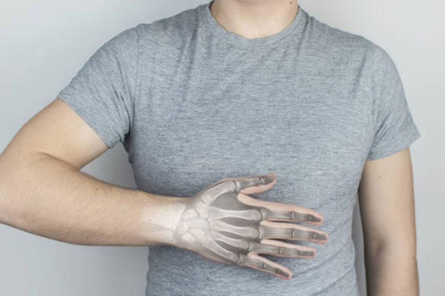
Scaphoid fractures of the wrist
A Scaphoid fracture is a fracture of one of the small bones of the wrist. This type of fracture often occurs after a fall on an outstretched hand. Symptoms of a Scaphoid fractures fracture usually include pain and tenderness in the area just below the base of the thumb. These symptoms may worsen when you try to pinch or hold something.
Treatment for a Scaphoid fractures fracture can range from orthosis to surgery, depending on the severity of the fracture and its location on the bone. Because parts of the Scaphoid fractures bone have a poor blood supply—and a fracture can further disrupt blood flow to the bone—complications of the healing process are common.
A reason
A boat fracture usually occurs when you fall onto an outstretched hand, with your weight landing on the palm of your hand. The end of the greater forearm bone (radius) may also break in this type of fall, depending on the position of the hand when descending.
Injury can also occur during sports activities or motor vehicle collisions.
Naval fractures occur in people of all ages, including children, and there are no specific risk factors or diseases that make you more likely to have a scaphoid fractures fracture. Some studies have shown that using wrist guards during high-energy activities such as snowboarding and snowboarding can help reduce your chance of breaking a bone around your wrist.
Symptoms
Scaphoid fractures usually cause pain and swelling in the anatomical snuff box and on the thumb side of the wrist. The pain may be severe when you move your thumb or wrist, or when you try to pinch or grip something.
Unless your wrist is deformed, it may not be obvious that your Scaphoid fractures bone is broken. With some canoe fractures, the pain is not severe and may be mistakenly thought of as a sprained wrist.
Pain in your wrist that doesn’t go away within a day of the injury could be a sign of a fracture – so it’s important to see a doctor if the pain persists. Prompt treatment of a scaphoid fracture will help to avoid potential complications.
Doctor’s Examination
Physical examination
During the examination, your doctor will talk to you about your general health and will ask you to describe your symptoms. The doctor will want to know how your injury occurred.
Your doctor will examine your wrist. With most fractures, there will be tenderness directly above the canoe in the anatomical snuff box. Your doctor will also look for:
- swelling
- bruising
- loss of movement
- the exams
X rays provide images of the dense structures, such as bones. Your doctor will order an X-ray to help determine if you have a scaphoid fracture and whether the broken pieces of bone have displaced. An X-ray will also help the doctor determine if you have any other fractures.
In some cases, a scaphoid fracture doesn’t show up on an X-ray right away. If your doctor suspects you have a fracture but it’s not visible on an X-ray, he or she may recommend that you wear a wrist splint or cast for 2 to 3 weeks and then come back for a follow-up X-ray. Often, scaphoid fractures become visible only on X-rays after a period of time. During this waiting period, you should wear a cast or cast and avoid activities that may cause further injury.
1. Magnetic resonance imaging (MRI)
Your doctor may order an MRI to learn more about the bones and the soft tissues in your wrist. An MRI can sometimes show a fractured canal before it can be seen on an X-ray.
2. Computerized tomography (CT) scan
A CT scan can be useful in detecting a fracture of the scaphoid bone and can also show whether the bones have displaced. Your doctor will use the information from the CT scan to help determine your treatment plan.
Treatment
The treatment your doctor recommends depends on a number of factors, including:
- The site of the fracture in bone
- Whether the bone fragments have been displaced
- Since when did you get injured?
Non-surgical treatment
A fracture near the thumb
Canoe fractures closest to the thumb (distal pole) usually heal within weeks with proper protection and restricted activity. This part of the scaphoid bone has a good blood supply, which is essential for healing.
For this type of fracture, your doctor may place your forearm and hand in a cast or splint. The cast or splint is usually just below the elbow and includes your thumb.
Recovery time varies from patient to patient
Your doctor will monitor your recovery with periodic X-rays or other imaging studies.
Fracture near the forearm
If the cane is broken in the middle of the bone (waist) or near the forearm (proximal pole), healing may be more difficult. These areas of the scapula do not have a very good blood supply.
If your doctor treats this type of fracture with a splint, the cast may include the thumb and extend over the elbow to help stabilize the fracture.
Bone stimulator
In some cases, your doctor may recommend the use of a bone stimulator to help fracture heal. This small device delivers low-intensity or pulsed electromagnetic waves that stimulate healing.
Surgical treatment
If the canoe is broken at the waist or proximal pole, or if pieces of bone are displaced, your doctor may recommend surgery. The goal of surgery is to realign and stabilize the fracture, giving it a better chance of healing.
Shorthand
During this procedure, your doctor will administer an anesthetic or anesthetic and put the bones back into their proper position. In some cases, this is done using a limited incision and special guided tools. In other cases, it is performed through an open incision with the direct manipulation of the fracture. For some fractures, your doctor may use a small camera called an “arthroscope” to help reduce them.
BY: Web Team
Magazine
COMMENTS: No Comments
 29 Mar 2022
Carpal Tunnel Syndrome
What is Carpal tunnel syndrome?
Carpal tunnel syndrome is a common condition that causes the pain, numbness, and tingling in the hand and arm. The condition occurs when one of the major nerves in the hand – the median nerve – is compressed as it travels through the wrist.
In most patients, the carpal tunnel syndrome gets worse over time, so early diagnosis and treatment are important. Early on, symptoms can often be relieved with simple measures such as wearing a wrist splint or avoiding certain activities.
Cause of Carpal tunnel syndrome
Most cases of the carpal tunnel syndrome are caused by a combination of factors. Studies show that women and the elderly are more likely to have this condition.
Other risk factors for the carpal tunnel syndrome include:
- Genetics. This is likely an important factor. The carpal tunnel may be smaller in some people or there may be anatomical differences that change the amount of nerve space – these features can run in families.
- Frequent manual use. Repeating the same hand and wrist movements or activities over an extended period of time may aggravate the wrist tendons, causing swelling that press on the nerve.
- Hand and wrist position. Doing activities that involve intense flexion or extension of the hand and wrist for an extended period of time can put more pressure on the nerve.
- The hormonal changes during pregnancy can cause swelling.
- Health conditions. Diabetes, rheumatoid arthritis, and thyroid imbalance are conditions associated with carpal tunnel syndrome.
Symptoms of Carpal Tunnel syndrome
Symptoms of carpal tunnel syndrome may include:
- Numbness, tingling, burning, and pain – primarily in the thumb, index, middle, and ring fingers
- Occasional bump-like sensations that radiate to the thumb, index, middle, and ring fingers
- The pain or tingling that may travel up the forearm toward the shoulder
- Weakness and clumsiness in the hand – this can make it difficult to perform precise movements such as buttoning your clothes
- dropping objects – due to weakness, numbness or loss of proprioception (perception of where your hand is in space)
In most cases, symptoms of carpal tunnel syndrome begin gradually – without specific injury. Many patients find that their symptoms come and go at the first. However, as the condition worsens, symptoms may occur more frequently or may last for longer periods of time.
Nocturnal symptoms are very common. Since many people sleep with the wrist bent, symptoms may wake you from sleep. During the day, symptoms often appear when holding something for an extended period of time with the wrist bent forward or backward, such as when using a phone, driving, or reading a book.
Treatment of Carpal Tunnel Syndrome
Although it is a gradual process, for most people, carpal tunnel syndrome will get worse over time without any form of treatment. For this reason, it is important to be evaluated and also diagnosed by your doctor early on. In the early stages, it may be possible to slow or stop progression of the disease.
1. Non-surgical treatment
If carpal tunnel syndrome is diagnosed and treated early, symptoms of carpal tunnel syndrome can often be relieved without surgery. If your diagnosis: is uncertain or if your symptoms are mild. The doctor will recommend the nonsurgical treatment first.
Non-surgical treatments may include:
Bracing or splinting
Wearing a brace or the splint at night will prevent you from bending your wrist while you sleep. Keeping your wrist straight or neutral reduces pressure on the nerve in the carpal tunnel. It may also be helpful to wear a splint during the day when doing activities that exacerbate symptoms.
Non-steroidal anti-inflammatory drugs (NSAIDs). Medicines such as ibuprofen and naproxen can help reduce pain and inflammation.
Activity changes
Symptoms often occur when your hand and wrist are in the same position for too long — particularly when your wrist is bent or extended.
If your job or recreational activities worsen your symptoms, changing or modifying these activities can help slow or stop the progression of the disease. In some cases, this may involve making the changes to your worksite or the workstation.
Steroid injections
Injecting a steroid into the carpal tunnel may relieve symptoms for a while.
Nerve gliding exercises
Some patients may benefit from exercises that help the median nerve to move more freely within the confines of the carpal tunnel.
Steroid injection
A corticosteroid, or cortisone, is a powerful anti-inflammatory agent that can be injected into the carpal tunnel. Although these injections often relieve painful symptoms or help calm a flare-up, sometimes their effect is temporary. Your doctor may also use cortisone shot to help diagnose carpal tunnel syndrome.
Surgical treatment
If nonsurgical treatment does not relieve symptoms after a while, your doctor may recommend surgery.
The decision to have surgery depends on the severity of your symptoms – how much pain and numbness you have in your hand. In long-term cases with persistent numbness and wasting of the thumb muscles, surgery may be recommended to prevent irreversible damage.
2. Surgical procedure
The surgical procedure performed for the carpal tunnel syndrome is called “carpal tunnel release.” There are the two different surgical ways to do this, but the goal is to relieve pressure on the median nerve by cutting ligament that forms the roof of the tunnel. This increases the size of the tunnel and reduces pressure on the median nerve.
In most cases, the carpal tunnel surgery is performed on an outpatient basis. The surgery can be performed under general anesthesia, which puts you to sleep, or under local anesthesia, which numbs only your hand and arm. In some cases, you’ll also be given a mild sedative through an intravenous (IV) line inserted into a vein in your arm.
Open carpal tunnel. In open surgery, your doctor makes a small incision in the palm of your hand and looks inside your hand and wrist through this incision. During the procedure, your doctor will split the transverse carpal ligament (the roof of the carpal tunnel). This increases the size of the tunnel and reduces pressure on the median nerve.
Thumbay University Hospital offers expert care through a multidisciplinary team of compassionate reproductive and caring professionals, perinatal specialists, diagnostic experts, nursing staff and highly trained technicians. Our team will assist you throughout your personal experience from diagnosis to your individual treatment plan.
BY: Web Team
Magazine
COMMENTS: No Comments
 28 Mar 2022
Adult Acne: Common Causes and Ways to Treat Them
Facing acne during adulthood is a common problem that a lot of people face. Normally acne can be a red bump on your skin. But, that’s just one form. In other cases, acne could be more severe involving cysts and pustules. Most adults are seen trying out everything from creams to serums to homemade remedies to cure their acne. But unfortunately, no product or treatment will work if you are clueless about the main cause of your acne breakouts.
The main reason behind the occurrence of acne is clogged pores. Pores that are present on your skin can get clogged as a result of dirt, oil, debris, bacteria, and skin flakes accumulating on the surface. Both men and women can have acne. For some, it can be mild, but for some, it could be severe.
What Causes the Breakouts?
As mentioned earlier, the basic cause of all acne is clogged pores. These pores are the openings surrounding your hair follicles and are also house to the sebaceous glands. These glands secrete sebum which is responsible for keeping the skin soft and protected. But when your pores get clogged, these glands won’t be able to secret sebum which will cause acne.
In some cases, following the simple step of CTM (Cleansing, Toning & Moisturizing) and exfoliating on a regular basis can help prevent acne to a great extent. But for others, it is quite complicated. The situation becomes quite frustrating when you try to figure out what is causing your acne.
Through this blog, we bring to you a list of some of the most common causes of acne and the best strategies to treat it.
Common Causes of Acne:
- Stress
According to the skin specialists in Ajman chronic stress is one of the most common causes of acne. When a person feels stressed, the adrenal gland releases cortisol otherwise known as the stress hormone. These hormones are present in our hair follicles too and other different types of skin cells. Though cortisol is responsible for a number of skin and hair-related issues, it is a very important compound present in the human body that helps regulate other processes in the body as well. These processes include the digestive system, immune system, and neurological systems. It is quite natural for the cortisol levels to fluctuate over time.
But when you feel too stressed, your body starts secreting too much cortisol which causes issues with the bodily processes and your skin. According to research, this hormone can create a favorable atmosphere for acne caused by bacteria.
- Wrong Products
There are a lot of products available in the market for skin-related issues for every type of skin. Though it is advised to be very careful about the products you apply to your skin irrespective of your skin type, this is even more important for people with oily skin.
If you have oily or combination skin, you should be using products that are ‘oil-free’, water-based, or non-comedogenic in nature. Using such products will reduce the chances of your pores getting clogged. You can also go for a gel-based moisturizer.
- Pollution
It might come as a surprise to you but the environment around you can have a severe impact on your skin, especially if it is acne-prone. The dirt and grime can settle on your skin and clog the pores which results in acne. But it is not just dirt that can cause skin issues. Ultra Violet (UV) rays can damage your skin too. Being exposed to the sun for long hours can cause skin discoloration, and cause fine lines, and aging. Also, it can make your skin too dry, leading to the production of excess oil to compensate for the dryness. And when there is excess oil, it can cause acne. Hence, our experienced skin specialists in Ajman will advise you to apply sunscreen (with at least SPF 30) to protect your skin from the harmful rays of the sun.
- Hormonal Changes
Fluctuation in hormones is another reason for acne. Do you often see the ‘red bump’ on your skin after a stressful day at work? Well…you will be surprised to know what hormonal changes can do to your skin.
For example, when your body produces progesterone (after ovulation), it increases the production of sebum in your skin which causes acne. Similarly, Androgens like testosterone can also ramp up the production of sebum causing hormonal acne.
Ways to Treat Adult Acne:
- Spot Treatments
Spot treatments are one of the best and fastest ways of treating acne. According to a skin specialist, a person suffering from severe acne should preferably use spot treatments containing benzoyl peroxide, which works great at eliminating the bacteria that cause acne. But people with sensitive skin need to be careful as the chemical can be harsh on their skin. In such a case, it is always advised to go for a patch test to be on the safer side.
- Exfoliators
Exfoliation is another important thing that people suffering from chronic acne should include in their daily skincare. Using exfoliators regularly can help prevent acne and treat it at the same time.
Whether you choose to go for a physical or a chemical exfoliant, rest assured that it will prevent breakouts by clearing out your pores and unclogging them. Glycolic acid is an ingredient that provides good results. That being said, you need to be careful not to exfoliate too often as it can cause irritation and make your skin dry and flaky. An experienced skin specialist will recommend exfoliating two to three times a week.
- Injecting Cortisone
In the case of cystic acne, there is a good chance that topical treatments (using salicylic acid, PHAs, benzoyl peroxide, etc.) might not work, And because they are big and painful zits deep into the skin, they can leave a scar if prodded. Hence, the best option is to drain it ASAP.
For draining cystic acne, you can get a shot of cortisone after consulting your dermatologist or you can apply a warm or a cold compress. Another way could be by applying a cream that contains hydrocortisone.
- Oral Medications
In certain cases, a dermatologist might recommend oral medications. Medicines that can manipulate hormones such as spironolactone are quite effective in controlling outbreaks in the chin and lower face areas that are caused by hormonal imbalances. Additionally, oral retinoids such as isotretinoin are considered powerful dermatological weapons against acne.
In case you are suffering from a severe case of adult acne and are looking for an experienced dermatologist in Ajman, contact the Thumbay University Hospital today to book an appointment with one of our specialists.
BY: Web Team
Magazine
COMMENTS: No Comments